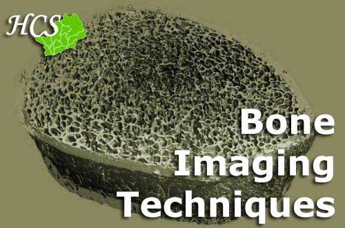
Bone Imaging Techniques
We have used novel scanning technology to look more closely at the structure of bone with the help of our HCS participants.
There are three types of scan we have performed in the HCS. The first, Dual X-ray Absorptiometry (DXA) allows us to look at bone thickness and to measure the proportions of bone, fat and muscle in the whole body. The second is peripheral Quantitative Computed Tomography (pQCT) which is a specialised, research scan. The images from this type of scan allow us to look more closely at bone, muscle and fat in arms and legs. The cousin of the pQCT is the high-resolution pQCT (HRpQCT). This scan provides such high quality images, that we can create a 3D model of the bone structure. It is so accurate that it has been called ‘a virtual bone biopsy’, meaning that we can tell as much about the structure of the bone from our scan, as you would be able to from looking at a sample of bone under a microscope.
We used the latter scanner, HRpQCT, to investigate the risk of fracturing in about 300 members of the 1930’s Hertfordshire birth cohort. We found that fractures can occur due to weakness of the cortex (outer shell of the bone) in some people, but can occur due to weakness of the trabeculae (core of bone) in others. Understanding that bones fracture due to weakness in different areas may mean that, in the future, we might be able to target different therapies to prevent fractures for different people depending on where the weakness is in their bones.
In another study we used DXA to analyse fat mass and lean (muscle) mass in a group of about 300 participants. We also used HRpQCT to study the internal structure of their bones. We found that higher fat mass was related to greater number and density of trabeculae (honey-comb structures within bone), whilst higher lean mass was related to increased area and thickness of the bone cortex (outer casing of bone). Our findings suggest that the proportions of fat and muscle tissues which make up our bodies can have effects on different parts of bone.
Reference List
Lu S, Fuggle NR, Westbury LD, Ó Breasail M, Bevilacqua G, Ward KA, Dennison EM, Mahmoodi S, Niranjan M, Cooper C. Machine learning applied to HR-pQCT images improves fracture discrimination provided by DXA and clinical risk factors. Bone. 2023 Mar;168:116653. doi: 10.1016/j.bone.2022.116653. Epub 2022 Dec 27.
Erratum in: Bone. 2024 May;182:117071. PMID: 36581259.
Fuggle NR, Westbury LD, Bevilacqua G, Titcombe P, Ó Breasail M, Harvey NC, Dennison EM, Cooper C, Ward KA. Level and change in bone microarchitectural parameters and their relationship with previous fracture and established bone mineral density loci. Bone. 2021 Jun;147:115937. doi:10.1016/j.bone.2021.115937. Epub 2021 Mar 22. PMID: 33766802; PMCID: PMC7611749.
Patel A, Edwards MH, Jameson KA, Ward KA, Fuggle N, Cooper C, Dennison EM. Longitudinal Change in Peripheral Quantitative Computed Tomography Assessment in Older Adults: The Hertfordshire Cohort Study. Calcif Tissue Int. 2018 Nov;103(5):476-482. doi: 10.1007/s00223-018-0442-0. Epub 2018 Jun 21. PMID: 29931460; PMCID: PMC6179140.
Paccou J, Edwards MH, Patsch JM, Jameson KA, Ward KA, Moss C, Dennison EM, Cooper C. Lower leg arterial calcification assessed by high-resolution peripheral quantitative computed tomography is associated with bone microstructure abnormalities in women. Osteoporos Int. 2016 Nov;27(11):3279-3287. doi: 10.1007/s00198-016-3660-1. Epub 2016 Jun 21. PMID: 27325126; PMCID: PMC5040512.
Paccou J, Ward KA, Jameson KA, Dennison EM, Cooper C, Edwards MH. Bone Microarchitecture in Men and Women with Diabetes: The Importance of Cortical Porosity. Calcif Tissue Int. 2016 May;98(5):465-73. doi: 10.1007/s00223-015-0100-8. Epub 2015 Dec 19. PMID: 26686695.
Paccou J, Edwards MH, Ward K, Jameson K, Moon R, Dennison E, Cooper C. Relationships between bone geometry, volumetric bone mineral density and bone microarchitecture of the distal radius and tibia with alcohol consumption. Bone.2015 Sep;78:122-9. doi: 10.1016/j.bone.2015.05.002. Epub 2015 May 8. PMID: 25959415.
Paccou J, Edwards MH, Ward KA, Jameson KA, Moss CL, Harvey NC, Dennison EM, Cooper C. Ischemic heart disease is associated with lower cortical volumetric bone mineral density of distal radius. Osteoporos Int. 2015 Jul;26(7):1893-901. doi: 10.1007/s00198-015-3132-z. Epub 2015 Apr 24. PMID: 25906240.
Dennison EM, Jameson KA, Edwards MH, Denison HJ, Aihie Sayer A, Cooper C. Peripheral quantitative computed tomography measures are associated with adult fracture risk: the Hertfordshire Cohort Study. Bone. 2014 Jul;64:13-7. doi: 10.1016/j.bone.2014.03.040. Epub 2014 Mar 27. PMID: 24680720.
Abdin-Mohamed M, Jameson K, Dennison EM, Cooper C, Arden NK; Hertfordshire Cohort Study Group. Volumetric bone mineral density of the tibia is not increased in subjects with radiographic knee osteoarthritis. Osteoarthritis Cartilage. 2009 Feb;17(2):174-7. doi: 10.1016/j.joca.2008.06.004. Epub 2008 Aug 5. PMID: 18684648.